Evaluation and Management of Precancerous and Atypical Skin Lesions
Actinic Keratoses and Dysplastic Nevi are common skin findings associated with sun exposure and individual skin risk factors. While these lesions are often benign or precancerous, careful dermatologic evaluation is important to identify lesions that may require treatment or monitoring.
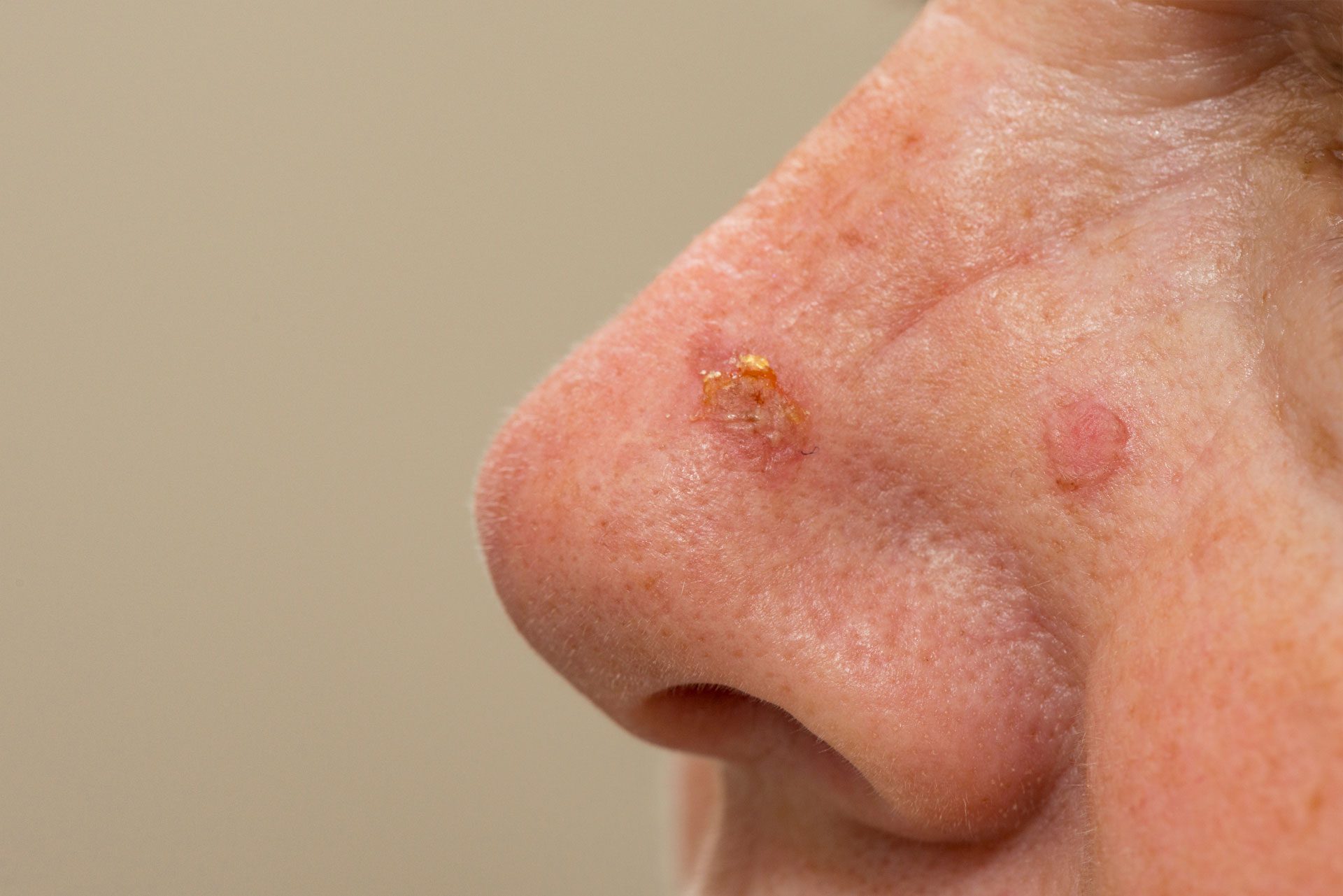
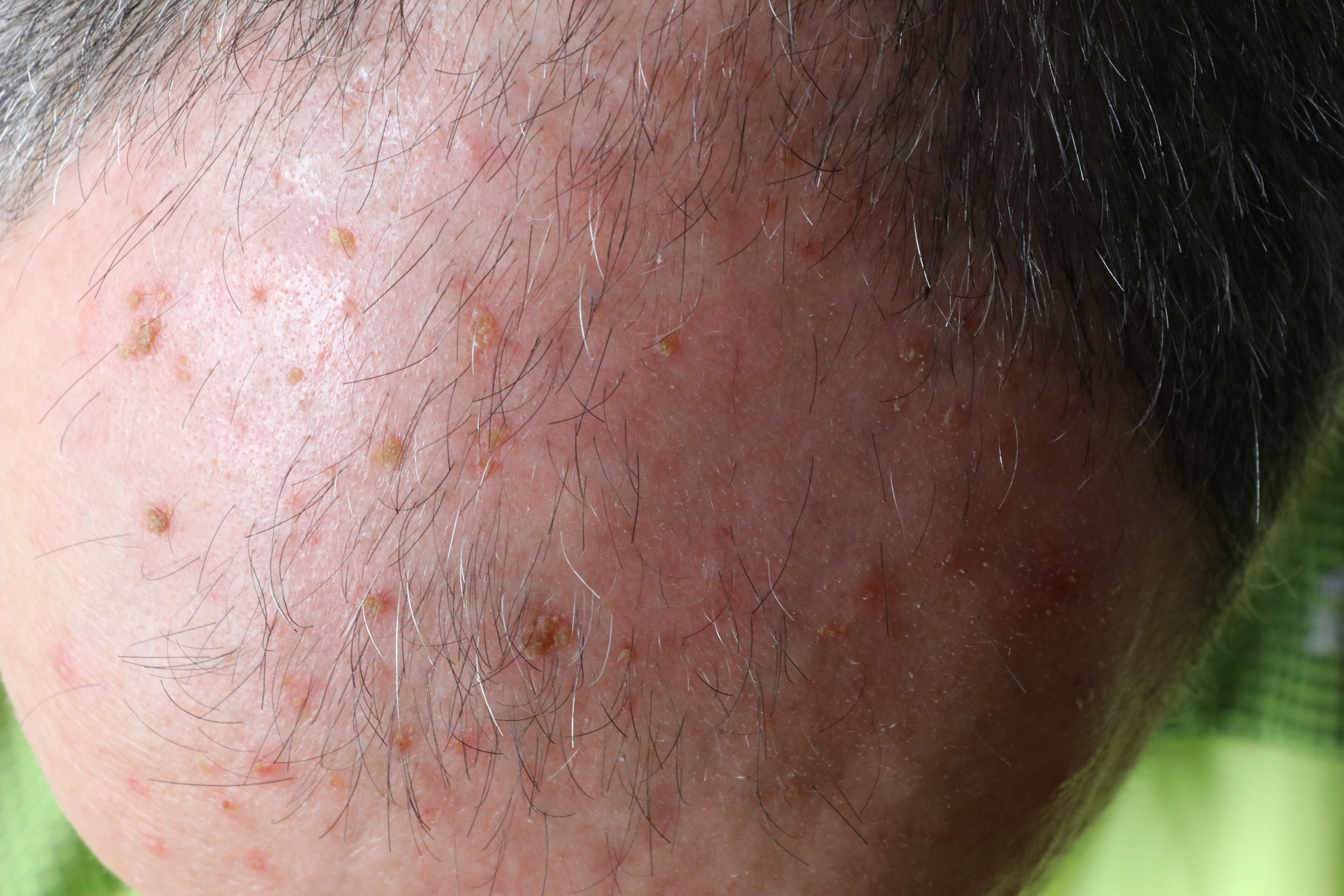
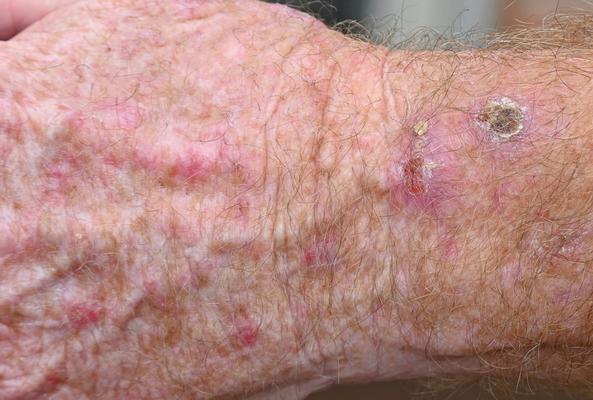
Actinic keratoses are rough, scaly patches that develop on sun-exposed areas of the skin, including the face, scalp, ears, arms, and hands. They are considered precancerous lesions and represent areas of cumulative sun damage.
Why Treatment of Actinic Keratoses are Important
Actinic keratoses have the potential to progress to squamous cell carcinoma (SCC). Studies suggest that:
Approximately 5–10% of actinic keratoses may progress to SCC over time
The risk is higher in patients with multiple lesions or extensive sun damage
Because it is not possible to predict which lesions will progress, treatment of actinic keratoses is recommended.
Field Cancerization
Actinic keratoses often occur in areas of field cancerization, where large areas of skin have sustained cumulative ultraviolet damage. This means:
Visible lesions represent only part of the damage
Surrounding skin may contain subclinical precancerous changes
New lesions may continue to develop over time
Field-directed treatments target both visible and subclinical lesions and are an important part of long-term skin cancer prevention.
Treatment of Actinic Keratosis
Treatment is based on the number, location, and extent of lesions.
Cryotherapy
Cryotherapy uses controlled freezing with liquid nitrogen to destroy individual actinic keratoses. This is commonly used for isolated lesions.
Photodynamic Therapy (PDT)
Photodynamic therapy is a light-activated treatment used to treat areas of sun-damaged skin affected by multiple actinic keratoses. This approach addresses field cancerization and helps reduce the risk of progression to squamous cell carcinoma.
Topical Therapies
Topical medications may be used to treat broader areas of sun damage and actinic keratoses. These treatments work over time to target abnormal cells within the skin.
Dysplastic Nevus (Atypical Mole)
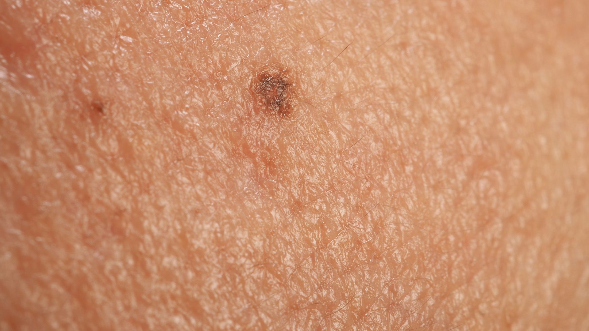
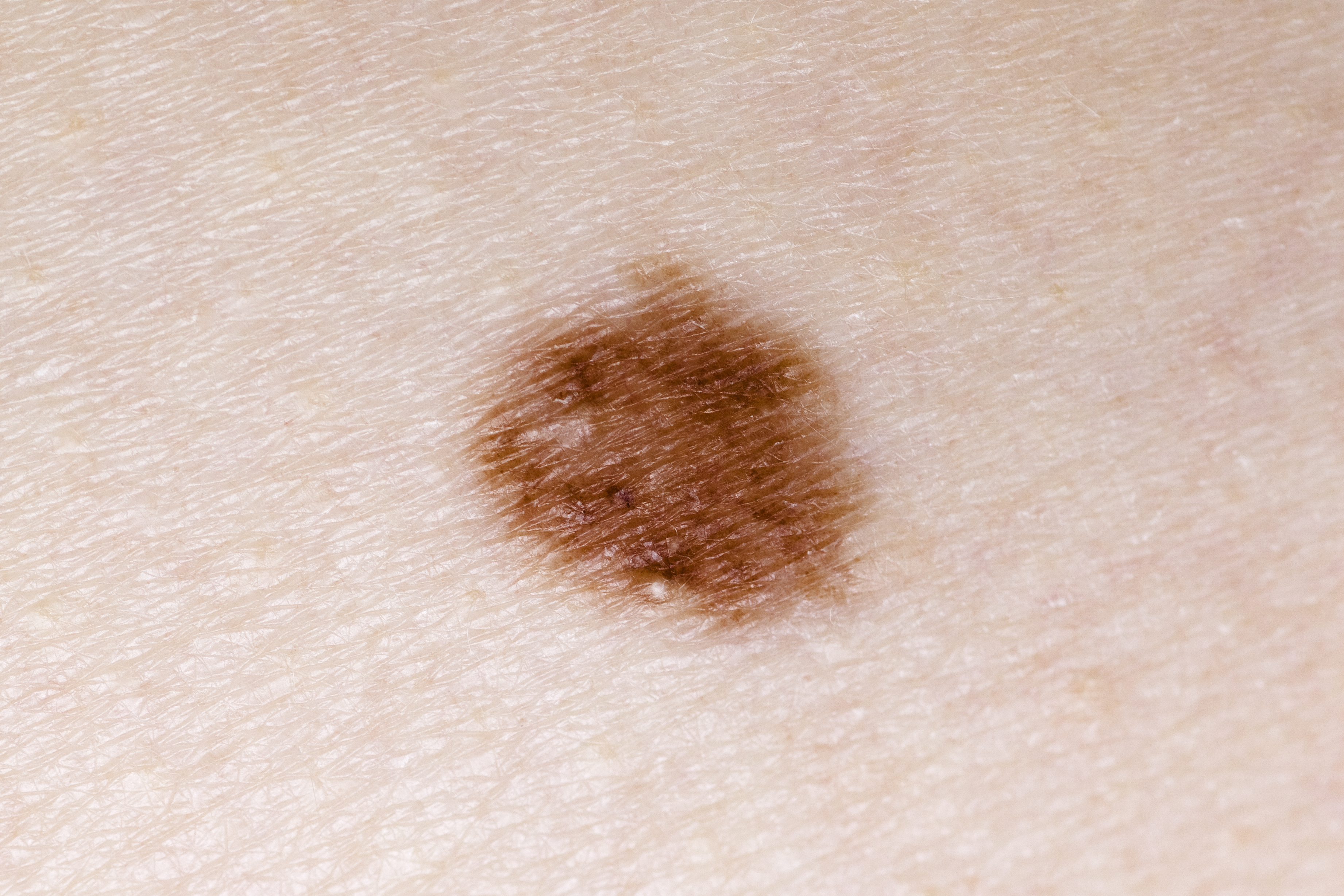
Dysplastic Nevi are atypical moles that may appear different from common moles. They often have irregular borders, variation in color, or larger size.
While Dysplastic Nevi are benign, they are considered markers of increased melanoma risk.
Why Dysplastic Nevi Matter
Individuals with dysplastic nevi may have:
An increased risk of melanoma
Multiple atypical moles that require monitoring
Lesions that may change over time
Careful evaluation helps distinguish benign atypical moles from lesions that may require biopsy.
Evaluation and Monitoring
Evaluation and monitoring are tailored to each patient’s individual risk profile and history.
Dermatologic Examination
A comprehensive skin examination includes evaluation of moles across the entire body. Particular attention is given to lesions that appear different from others.
Dermoscopy
Dermoscopy may be used to evaluate pigment patterns and structures not visible to the naked eye.
Biopsy
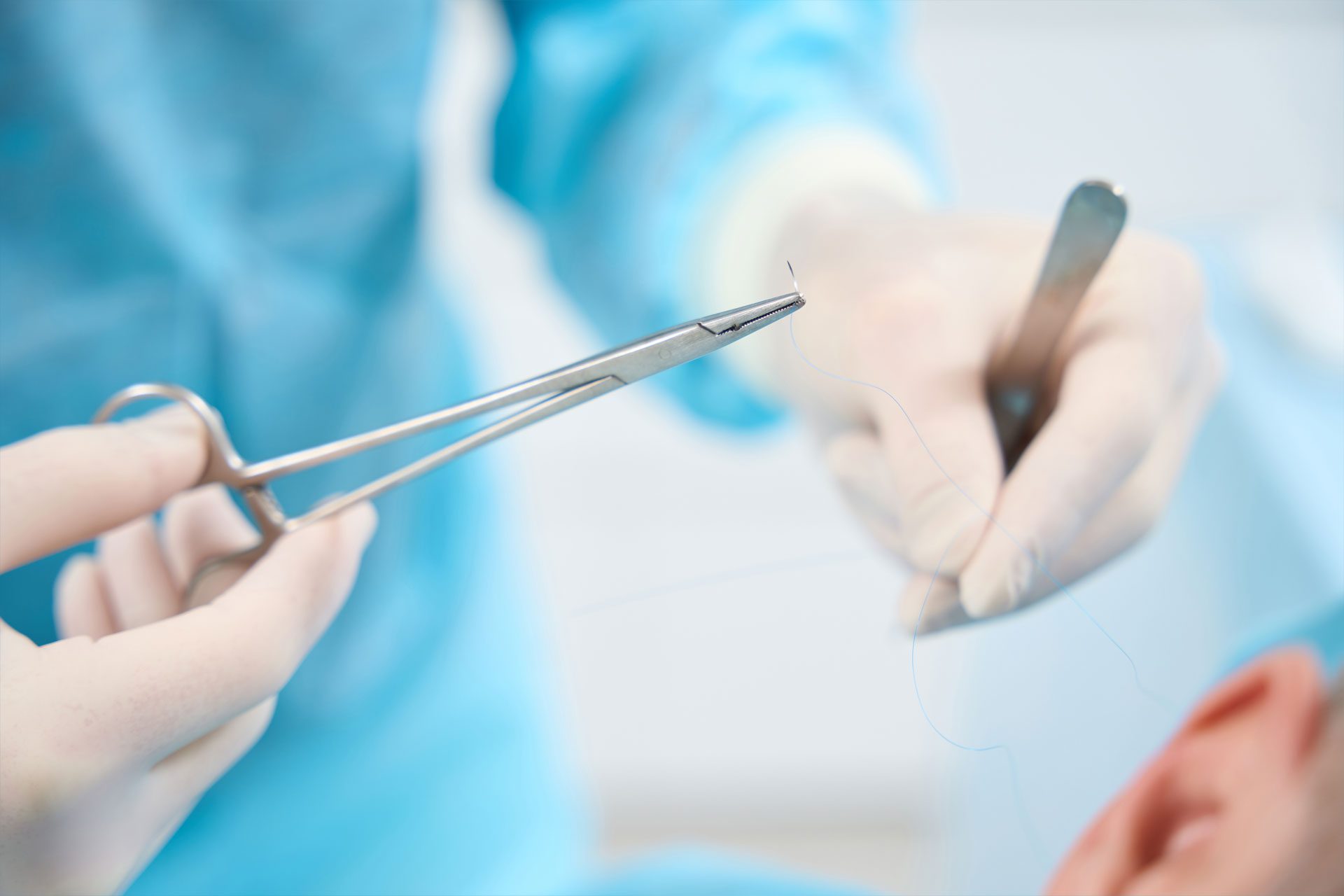
If a mole appears suspicious or shows concerning changes, a biopsy may be performed to confirm the diagnosis.
Ongoing Surveillance
Patients with actinic keratoses or dysplastic nevi benefit from ongoing dermatologic surveillance.
Follow-up care may include:
Regular total body skin examinations
Monitoring for new or changing lesions
Individualized follow-up intervals based on risk factors
Prevention and Skin Health
Preventive measures play an important role in managing both actinic keratoses and dysplastic nevi.
Recommendations include:
Daily use of sunscreen and sun protection
Avoidance of excessive ultraviolet (UV) exposure
Routine skin examinations
Monitoring moles and skin lesions for change
When to Seek Evaluation
Evaluation is recommended for:
Rough, scaly lesions that persist
New or changing moles
Lesions that bleed, itch, or do not heal
Any mole that appears different from others
Schedule a Skin Examination
Early evaluation and treatment of actinic keratoses and careful monitoring of dysplastic nevi are important components of skin cancer prevention.